

In many cases, our in-house laboratory can offer great insight to help your pet’s vet confirm a diagnosis. However, there are times where an external laboratory can provide more comprehensive testing. In these cases, it’s worth the wait!


Anesthesia is not usually needed for most ultrasound examinations, unless biopsies are to be taken. The technique is totally painless and most pets will lie comfortably while the scan is being performed. Occasionally, if the pet is very frightened or worried, a sedative may be necessary.
In most cases, the fur must be shaved to perform an ultrasound examination. Since ultrasound waves are not transmitted through air, it is imperative that the probe makes complete contact with the skin. In some cases, such as pregnancy diagnosis, it may be possible to get adequate images by moistening the hair with rubbing alcohol and applying a copious amount of water-soluble ultrasound gel.
Since an ultrasound study is performed in real time, the results of what is seen are known immediately. In some cases, the ultrasound images may be sent to a veterinary radiologist for further consultation. When this occurs, the final report may not be available for a few days.
We’re delighted to be able to offer this important diagnostic tool to you and your pet. Please feel free to call us if you have additional questions about ultrasound!
The ultrasound waves are converted into an image that is displayed on the monitor, giving a 2-dimensional “picture” of the tissues under examination. The technique is invaluable for the examination of internal organs and is very useful in the diagnosis of cysts and tumors.

Ultrasound
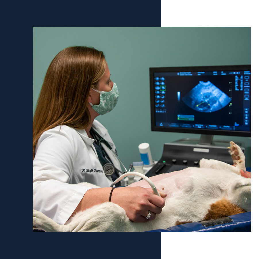
An ultrasound examination, also known as ultrasonography, is a non-invasive imaging technique that allows internal body structures to be seen by recording echoes or reflections of ultrasonic waves. Ultrasound equipment directs a narrow beam of high frequency sound waves into the area of interest.
The ultrasound waves are converted into an image that is displayed on the monitor, giving a 2-dimensional “picture” of the tissues under examination. The technique is invaluable for the examination of internal organs and is very useful in the diagnosis of cysts and tumors.
Anesthesia is not usually needed for most ultrasound examinations, unless biopsies are to be taken. The technique is totally painless and most pets will lie comfortably while the scan is being performed. Occasionally, if the pet is very frightened or worried, a sedative may be necessary.
In most cases, the fur must be shaved to perform an ultrasound examination. Since ultrasound waves are not transmitted through air, it is imperative that the probe makes complete contact with the skin. In some cases, such as pregnancy diagnosis, it may be possible to get adequate images by moistening the hair with rubbing alcohol and applying a copious amount of water-soluble ultrasound gel.
Since an ultrasound study is performed in real time, the results of what is seen are known immediately. In some cases, the ultrasound images may be sent to a veterinary radiologist for further consultation. When this occurs, the final report may not be available for a few days.
We’re delighted to be able to offer this important diagnostic tool to you and your pet. Please feel free to call us if you have additional questions about ultrasound!


